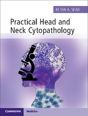
Practical Head and Neck Cytopathology
with Online Static Resource
AUD$114.95 inc GST
- Author: Ketan A. Shah, John Radcliffe Hospital, Oxford
- Date Published: February 2015
- availability: Available
- format: Print/online bundle
- isbn: 9781107443235
AUD$
114.95
inc GST
Print/online bundle
Looking for an inspection copy?
Please email [email protected] to enquire about an inspection copy of this book
-
Practical Head and Neck Cytopathology provides an engaging and holistic approach to the cytologic diagnosis of head and neck masses, covering both the technique of needle aspiration and its microscopic interpretation. A suitable sample is critical for arriving at the diagnosis and this book provides organ-specific tips on the FNA technique. Presence of different organs and structures in the head and neck region, as well as morphologic overlap amongst disparate entities, adds to the complexity of cytologic diagnosis. This book presents an easy-to-follow, stepwise approach to diagnosis, using concise bulleted text to highlight key features. Common and infrequently encountered mimics are included and interpretative challenges are discussed, facilitating a robust diagnosis. Numerous high-quality photomicrographs aid recognition of both common and rare features. A valuable resource for trainee and practising pathologists alike, this book is based on universally-used techniques and routinely prepared material, which are relevant to cytopathologists across the globe.
Read more- Provides a practical and holistic approach to the diagnosis of head and neck masses, including descriptions of key clinical features, practical tips on the aspiration technique, a stepwise guide to cytologic assessment, and discussion of diagnostic challenges
- Easy-to-follow, bulleted text provides succinct descriptions, emphasizing individual characteristics and cytologic features
- Numerous detailed photomicrographs illustrate classical features and variations in morphology, complementing the text and aiding recognition of entities
Customer reviews
Not yet reviewed
Be the first to review
Review was not posted due to profanity
×Product details
- Date Published: February 2015
- format: Print/online bundle
- isbn: 9781107443235
- length: 240 pages
- dimensions: 255 x 195 x 20 mm
- weight: 0.97kg
- contains: 675 colour illus. 6 tables
- availability: Available
Table of Contents
Preface
Acknowledgement
1. Aspiration technique and stains
2. Lymph nodes
2.1. Introduction
2.2. Reactive lymph node hyperplasia
2.3. Granulomatous inflammation
2.4. Lymphoma
2.5. Metastatic disease
2.6. Miscellaneous lymph node conditions
2.7. Approach to lymph node cytology
3. Salivary glands
3.1. Introduction and cytology of normal salivary glands
3.2. Proposed reporting system of salivary gland aspirates
3.3. Inflammatory salivary gland lesions
3.4. Cystic salivary gland lesions
3.5. Benign salivary gland neoplasms
3.6. Basal cell neoplasms
3.7. Oncocytic lesions
3.8. Malignant salivary gland neoplasms
3.9. Approach to salivary gland cytology
3.10. Role of repeat FNA
4. Thyroid
4.1. Introduction
4.2. Colloid goiter
4.3. Thyroiditis
4.4. Papillary carcinoma
4.5. Follicular neoplasms
4.6. Medullary carcinoma
4.7. Poorly differentiated carcinoma
4.8. Undifferentiated (anaplastic) carcinoma
4.9. Hyalinizing trabecular tumour
4.10. Lymphoma
4.11. Approach to thyroid cytology
5. Cystic neck lesions
5.1. Introduction
5.2. Branchial cyst
5.3. Thyroglossal cyst
5.4. Lymphangioma
5.5. Seroma and lymphocele
6. Other neck lesions
6.1. Introduction
6.2. Abscess
6.3. Cervicofacial actinomycosis
6.4. Parathyroid lesions
6.5. Suture granuloma
6.6. Lipoma
6.7. Spindle cell lipoma
6.8. Nodular fasciitis
6.9. Paraganglioma
6.10. Pilomatricoma
6.11. Schwannoma
Index.
Sorry, this resource is locked
Please register or sign in to request access. If you are having problems accessing these resources please email [email protected]
Register Sign in» Proceed
You are now leaving the Cambridge University Press website. Your eBook purchase and download will be completed by our partner www.ebooks.com. Please see the permission section of the www.ebooks.com catalogue page for details of the print & copy limits on our eBooks.
Continue ×Are you sure you want to delete your account?
This cannot be undone.
Thank you for your feedback which will help us improve our service.
If you requested a response, we will make sure to get back to you shortly.
×
