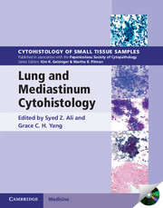
Lung and Mediastinum Cytohistology
with CD-ROM
Part of Cytohistology of Small Tissue Samples
- Editors:
- Syed Z. Ali, The Johns Hopkins University School of Medicine
- Grace C. H. Yang, Weill Medical College of Cornell University
- Date Published: May 2012
- availability: Available
- format: Mixed media product
- isbn: 9780521516587
Mixed media product
Looking for an examination copy?
This title is not currently available for examination. However, if you are interested in the title for your course we can consider offering an examination copy. To register your interest please contact [email protected] providing details of the course you are teaching.
-
Each volume in this richly illustrated series, sponsored by the Papanicolaou Society of Cytopathology, provides an organ-based approach to the cytological and histological diagnosis of small tissue samples. Benign, pre-malignant and malignant entities are presented in a well-organized and standardized format, with high-resolution color photomicrographs, tables, tabulated specific morphologic criteria and appropriate ancillary testing algorithms. Example vignettes allow the reader to assimilate the diagnostic principles in a case-based format. This volume provides comprehensive coverage of lung and mediastinal cytopathology. It presents a correlation of findings from fine-needle aspiration and exfoliative cytology and histologic findings obtained via core needle biopsies and surgical specimens. With a focus on malignant tumors, the full spectrum of inflammatory disorders, infectious diseases, hyperplasias and benign tumor or tumor-like lesions are also covered in detail. With over 500 printed photomicrographs and a CD-ROM offering all images in a downloadable format, this is an important resource for all anatomic pathologists.
Read more- Covers findings from the light microscope, correlative radiological findings, and immunohistochemical, genetic, molecular and other diagnostic modalities
- Richly illustrated with over 500 high-quality colour images, and a CD-ROM contains all images in a downloadable format
- Discusses the full spectrum of inflammatory disorders, infectious diseases and hyperplasias, with a focus on malignant tumors
Customer reviews
Not yet reviewed
Be the first to review
Review was not posted due to profanity
×Product details
- Date Published: May 2012
- format: Mixed media product
- isbn: 9780521516587
- length: 290 pages
- dimensions: 284 x 223 x 17 mm
- weight: 1.28kg
- contains: 500 colour illus. 20 tables
- availability: Available
Table of Contents
Preface
1. Introduction to lung cytopathology and small tissue biopsy Paul E. Wakely, Jr and Raheela Ashfaq
2. Normal anatomy, histology and cytology Andrea Subhawong
3. Infectious diseases Joseph D. Jakowski and Celeste N. Powers
4. Other non-neoplastic lesions Stefan E. Pambuccian
5. Benign lung tumors and tumor-like lesions Grace C. H. Yang
6. Squamous, large cell, and sarcomatoid carcinomas Yener S. Erozan and Grace C. H. Yang
7. Adenocarcinoma Jun Zhang and Grace C. H. Yang
8. Neuroendocrine neoplasms Momin T. Siddiqui
9. Uncommon primary neoplasms Armanda Tatsas and Syed Z. Ali
10. Metastatic and secondary neoplasms Christopher Owens
11. Anterior mediastinum Oscar Lin and Andre L. Moreira
12. Middle and posterior mediastinum Hong Q. Peng and Grace C. H. Yang
13. Role of ancillary studies Srinivas R. Mandavilli and Richard W. Cartun
Index.
Sorry, this resource is locked
Please register or sign in to request access. If you are having problems accessing these resources please email [email protected]
Register Sign in» Proceed
You are now leaving the Cambridge University Press website. Your eBook purchase and download will be completed by our partner www.ebooks.com. Please see the permission section of the www.ebooks.com catalogue page for details of the print & copy limits on our eBooks.
Continue ×Are you sure you want to delete your account?
This cannot be undone.
Thank you for your feedback which will help us improve our service.
If you requested a response, we will make sure to get back to you shortly.
×
