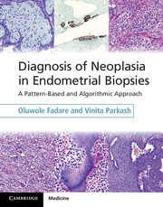
Diagnosis of Neoplasia in Endometrial Biopsies
A Pattern-Based and Algorithmic Approach
Book and Online Bundle
- Authors:
- Oluwole Fadare, Vanderbilt University, Tennessee
- Vinita Parkash, Yale University, Connecticut
- Date Published: September 2014
- availability: Available
- format: Mixed media product
- isbn: 9781107040434
Mixed media product
Looking for an inspection copy?
This title is not currently available for inspection. However, if you are interested in the title for your course we can consider offering an inspection copy. To register your interest please contact [email protected] providing details of the course you are teaching.
-
With its unique algorithmic and pattern-based approach, Diagnosis of Neoplasia in Endometrial Biopsies is an essential practical guide to interpreting endometrial biopsy samples. All potential entities are classified based on the dominant histologic pattern, with each resulting sub-group progressively sub-classified to reach a diagnosis. Decision tree flowcharts facilitate rapid narrowing of the differential diagnosis. Recent advancements are discussed and explained, and strengths and limitations of diagnostic tests are identified in the context of their application to the biopsy sample. Lavishly illustrated throughout, this book serves the practising pathologist as a scope-side assistant for quick reference, up-to-date guidance, and recommendations for ancillary testing. For the resident, this book facilitates quick and comprehensive mastery of the interpretation and diagnosis of endometrial biopsies. The book is packaged with a password, giving the user online access to all the text and images.
Read more- Over 400 high-quality color images, including over 250 taken from biopsy cases, enabling easy identification of key features as they pertain to biopsy samples
- Describes a pattern-based approach to the diagnosis of neoplasms in endometrial biopsies and curettages, facilitating rapid narrowing of differential diagnosis
- Offers up-to-date information with a practical algorithmic format, providing a quick and easy reference
Reviews & endorsements
'The book's tone is both descriptive and analytical. Given the title, every chapter has a plethora of flow charts and photomicrographs which, if indeed adhered to, will make life much easier for histopathologists reporting endometrial biopsies … [This book] should be considered as a valuable addition to the increasing number of bench books on uterine pathology.' The Bulletin of the Royal College of Pathologists
Customer reviews
Not yet reviewed
Be the first to review
Review was not posted due to profanity
×Product details
- Date Published: September 2014
- format: Mixed media product
- isbn: 9781107040434
- length: 182 pages
- dimensions: 282 x 222 x 15 mm
- weight: 0.9kg
- contains: 393 colour illus. 15 tables
- availability: Available
Table of Contents
Preface
1. General principles in the evaluation of endometrial samples
2. Endometrial samples with roughly equal ratio of glands to stroma
3. Lesions with epithelium to stroma ratio in excess of 1:1
4. Purely epithelial proliferations (no significant stromal component)
5. Spindle cell and myxoid lesions
6. Round cell lesions
7. Epithelioid cell lesions
8. Trophoblastic and gestational lesions
Index.
Sorry, this resource is locked
Please register or sign in to request access. If you are having problems accessing these resources please email [email protected]
Register Sign in» Proceed
You are now leaving the Cambridge University Press website. Your eBook purchase and download will be completed by our partner www.ebooks.com. Please see the permission section of the www.ebooks.com catalogue page for details of the print & copy limits on our eBooks.
Continue ×Are you sure you want to delete your account?
This cannot be undone.
Thank you for your feedback which will help us improve our service.
If you requested a response, we will make sure to get back to you shortly.
×
